How is Ocular Rosacea Diagnosed?
The diagnosis of ocular rosacea is made by taking a thorough medical history, listening to your symptoms and examining your eyes with a biomicroscope (slit lamp). Special attention is paid to evaluating your eyelids, eyelid margins, eyelashes, and meibomian gland orifices (openings). Gentle digital pressure is applied to your eyelids to assess the quality and quantity of the oils secreted by your meibomian glands which reflects the functioning of your Meibomian glands.
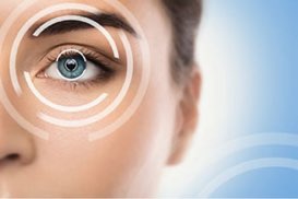
The gold standard to directly evaluate your meibomian glands structurally and anatomically is to image them using advanced technology known as LipiScan. LipiScan is a meibographer which captures images of your meibomian glands. Using this state-of-the-art technology your meibomian glands can be viewed to assess and quantify them structurally.
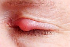
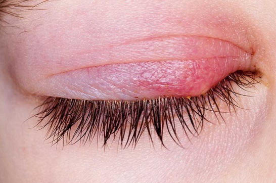
When you have either functional or structural issues with your meibomian glands this is known as meibomian gland dysfunction, or MGD. Blepharitis, meibomian gland dysfunction, and ocular rosacea are extremely closely related conditions which, to a large extent, have the same treatment protocols.

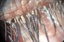
The Meibomian glands appear as white vertical lines in the eyelids in the photos below:


Once your eyes and eyelids have been evaluated, depending upon the degree to which ocular rosacea is an issue for you, Dr. Muller will discuss your treatment options. Dr. Muller's suggestions can range from homeopathic/natural treatments, to treatments involving medications and mechanical cleansing.