What is Neurotrophic Keratitis?
Neurotrophic keratitis is a problem with the surface of the eye resulting from a decrease in corneal sensitivity caused by damage to the corneal nerves, or the nerves surrounding and supplying the surface of the eye.
Neurotrophic keratitis is far more common than previously reported. The incidence of neurotrophic keratitis is underreported in part due to the lack of routine clinical testing of corneal sensitivity.
Dr. Muller evaluates corneal sensitivity on every patient she examines.
Like most other medical conditions neurotrophic keratitis presents, and should be thought of, as a spectrum of disease. Corneal hypoesthesia, or a decreased corneal sensation, can range from mild, to moderate, to severe.
Why are the corneal nerves important; what do they do?
First, the corneal nerves play an important sensory role in reflex tearing which protects the surface of the eye. An example of this would be if a gust of wind blows a particle (debris) into your eye, the sensory nerves on the surface of your eye will feel it and reflexively you will start tearing in an effort to wash out the foreign body. This is your eye’s way of protecting itself.
Second, we all have transparent skin on the surface of our eyes (called epithelium). The corneal nerves play an integral role in supporting the health, growth and repair of this skin.
What happens when your corneal nerves are impaired?
When your corneal nerves are damaged, as in neurotrophic keratitis, the overall health of the surface of your eye declines due to the impairment of the ability of the skin on the front of your eye to heal properly. This can result in the skin on the surface of your eyes becoming more friable and breaking down.
As the surface of your eye breaks down and begins to develop irregularities, including cracks and breaks, you may experience blurry, or fluctuating vision.
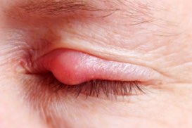
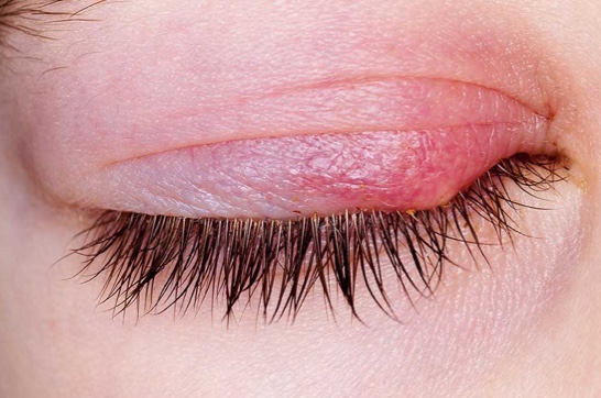
The cracks and breaks (open wounds) on the surface of your eye can range from small openings, known as superficial punctate keratitis (SPK), to recurrent and/or persistent corneal epithelial defects. These open wounds can lead to thinning of your cornea, scarring and even corneal perforation. The open wounds can also become infected (known as ulcers).
Prognostic indicators in neurotrophic keratitis include the degree of sensory loss, the duration of the condition, and the presence of other ocular surface issues which cause additional irritation and inflammation of the cornea such as dry eye, environmental allergies, meibomian gland dysfunction, blepharitis, ocular rosacea, demodex, contact lens related ocular irritation and inflammation, other.
People with reduced corneal sensitivity must be educated and instructed to seek proper medical evaluation immediately if their eye(s) become red, irritated, or they experience any changes in their vision. People with damage to their nerves may not feel as much pain or irritation as someone who doesn’t have this reduced sensitivity; they therefore must be cognizant of lowering the threshold for which they seek medical attention.
What causes Neurotrophic Keratitis?
- Contact lens wear/over wear
- Chronic use of eye drops containing preservatives
- Long term use of certain topical medications (i.e. Timolol, Sulfacetamide, Betaxolol, Diclofenac sodium, Ketorolac, etc.)
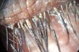
- Chronic irritation and inflammation associated with dry eye, environmental allergies, ocular rosacea, blepharitis, demodex and/or meibomian gland dysfunction (MGD)
- Herpetic infections of the cornea (simplex or zoster)
- Prior surgery which has damaged the nerves in this area (i.e. for trigeminal neuralgia, acoustic neuroma, other)
- Systemic diseases that can cause neurotrophic keratitis include diabetes, multiple sclerosis, and vitamin A deficiency
- Previous eye surgery (i.e. laser, cataract and/or glaucoma surgery)
- The normal aging process
- Other less common causes
Symptoms of Neurotrophic Keratitis
If you have neurotrophic keratitis, because of your reduced corneal sensitivity due to the damage to your corneal nerve endings you may have mild, or no symptoms at all. Alternatively, you can experience any of the following:
- Stinging, burning or irritation of your eyes
- Decreased, blurry or fluctuating vision
- Mucus discharge from your eyes
- Sensitivity to light (photophobia)
- Redness
- A “foreign body sensation”, as if you have an eyelash or a grain of sand in your eye(s)
- Discomfort wearing contact lenses
How is Neurotrophic Keratitis Diagnosed?
The diagnosis of neurotrophic keratitis is made by taking a careful ocular, medical, and surgical history, performing a thorough examination of the surface of your eyes using a biomicroscope with various medical dyes, and formally assessing your corneal sensitivity.
Treatment of Neurotrophic Keratitis:
The treatment of neurotrophic keratitis is based upon the severity of your condition and is aimed at maintaining the health of the surface of your eyes. Concomitant ocular issues causing additional irritation and inflammation such as dry eye, environmental allergies, ocular rosacea, blepharitis, meibomian gland dysfunction, herpes infections, and contact lens associated irritation and inflammation, should also be addressed.
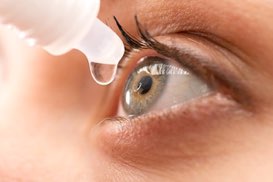
- Topical lubrication with preservative-free artificial tears, gels, and ointments
- Punctal plugs
- Low dose oral doxycycline or tetracycline (which have shown anti-inflammatory and anti-collagenolytic activity)
- Prescription anti-inflammatory eye drops
- Autologous serum
- Amniotic membranes - New York Times Article Regarding the Medical Use of Placentas and Amniotic Membranes (October 8, 2024)
- Exosome Therapy
- Oxervate (Cenegermin) is an FDA approved topical recombinant nerve growth factor. It is an eye drop which is instilled in the affected eye(s) 6 times each day at 2-hour intervals for 8 weeks.
- Scleral contact lenses
- Compounded topical insulin in the form of eye drops is an emerging area of research showing promise in treating dry eye disease, Meibomian gland dysfunction, ocular rosacea and blepharitis, collectively known as ocular surface diseases (OSD). These conditions involve inflammation, dryness, and damage to the cornea and conjunctiva (the ocular surface), lacrimal and Meibomian glands. Insulin has been found to have beneficial effects due to its anti-inflammatory and tissue-repairing properties, its ability to stimulate tear production and to improve corneal nerve function.Insulin eye drops are believed to promote cell growth and repair of the surface of the eyes, the lacrimal glands and the Meibomian glands, and to accelerate corneal healing, especially in cases of persistent epithelial defects (PEDs) and neurotrophic keratitis (NK). Additional studies suggest topical insulin can help restore corneal nerve function, which is crucial to supporting the health of the cornea and ocular surface.
Learn more by visiting: this page.