By Wendy Lyons Sunshine
Ophthalmology Management, Volume: 20, Issue: August 2016, page(s): 60, 63, 64
A surprising number of patients with a compromised tear film never actually complain of dryness. How is that possible?
“We used to think that dry eye was just a tear deficiency,” says Anat Galor, MD, MSPH, a staff physician at the Miami Veterans Administration Medical Center, and associate professor of clinical ophthalmology at Bascom Palmer Eye Institute. “Now we know it is a much more complicated pathophysiology.”
Patients might not grumble about dryness but still could have poor ocular surfaces. Others can complain of symptoms that are seemingly unrelated to dryness or they might only occur in specific situations.
As researchers continue to expand their understanding of the constellation of syndromes known as dry eye disease (DED), new insights emerge. Now clinicians realize that systemic disease, medications, environmental factors and nerve damage can all lead to “asymptomatic dry eye.”
“Dry eye should always be assessed in patients who are at risk, even if they have no dryness or discomfort symptoms,” says Esen K. Akpek, MD, associate director at the Jerome L. Greene Sjögren’s Syndrome Center at Johns Hopkins, and director of the Ocular Surface Diseases Clinic of The Wilmer Eye Institute.
So how do clinicians go about unmasking this “silent” dry eye?
Tip-offs to asymptomatic DED
When patients complain of fluctuating vision — especially while reading, or if they have trouble tolerating contact lenses or feel like they need to blink more often to see clearly — those could be clues of asymptomatic dry eye. Some patients complain that their eyes “feel tired” or that they’re re-reading the same sentence over and over. Others develop an awareness of their eyes after a long airplane flight.
“Patients often present with a chief complaint of blurred vision, especially when reading or at the computer,” says Alice T. Epitropoulos, MD, FACS, at The Eye Center of Columbus, and clinical assistant professor at Ohio State University Wexner Medical Center.
The most vulnerable patients who have dry eye but not the dryness complaint are those with diabetes or who are on glaucoma medications, says Dr. Akpek. “Also, in advanced cases of dryness, such as patients with Sjögren’s Syndrome, discomfort symptoms could be minimal due to changes in corneal nerve plexus. Yet when you stain the ocular surface there might be intense staining, which can have a huge impact on vision.”
A complete medical history offers important clues. Nerve damage will prevent complaints — and put these patients at higher risk, because it is unusual to address dry eye in asymptomatic patients. Other potential related culprits can include LASIK or cataract surgery, with their iatrogenic corneal nerve damage, and systemic conditions such as rheumatoid arthritis, lupus or thyroid disease.
Jay S. Pepose, MD, PhD, FARVO, a professor of clinical ophthalmology at Washington University School of Medicine, points out that tear hyperosmolarity leads to the loss of corneal epithelial cell microvilli and glycocalyx. “That reduces these cells’ ability to bind mucin and water, leading to cell death, exposure of corneal nerves and damage to the ocular surface.”
“So many systemic conditions show up with eye manifestations,” concurs Dr. Galor. She says a dry ocular surface can result from rheumatological syndromes and many chemotherapies, particularly hormone treatments for prostate cancer. Dr. Galor has participated in research that even found links between dry eye and depression and anxiety.1 “The ophthalmologist needs to continuously work with patients to figure out which other problems are affecting dry eye.”
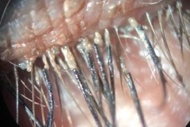
Even cosmetic practices are a risk. For example, the patient may be sleeping in their contact lenses or not cleaning them properly. Cynthia Matossian, MD, of Matossian Eye Associates, says, “I look for or ask about lash enhancements, lash embellishments, or false lashes — all of which can lead to ocular surface disease that can be asymptomatic initially.”
Finding the asymptomatic patient
A careful screening — especially of surgical patients — is important in identifying the asymptomatic patient. Clinicians should look for high osmolarity, diabetes, herpes zoster, cancer treatment, LASIK and prior systemic conditions that impact the ocular surface. A complete medical history will reveal issues that deplete physiologic reserves and increase corneal desensitization.
Dr. Epitropoulos uses a dry eye questionnaire and asks technicians to watch for symptoms of burning, tearing, grittiness and fluctuations in vision. These symptomatic patients receive point-of-care (POC) testing including tear osmolarity and matrix metalloproteinase-9 (MMP-9) prior to drops. “This is particularly important to diagnose in our cataract patients since dry eye can affect the accuracy of topography and keratometry and subsequent IOL power calculations,” she says.
“Even in asymptomatic cases, at the very least corneal staining with fluorescein should be assessed as part of a routine slit-lamp exam,” says Dr. Akpek.
Dr. Pepose concurs. “Some asymptomatic patients, particularly with advanced disease, can show signs such as staining of the ocular surface with vital dyes, such as lissamine green, and punctate staining of the cornea with fluorescein.”
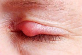
He reminds that lid disease can also be at play. “Meibomian gland inspissation can be assessed by expression and meiboscopy, leading to evaporative dry eye. Tear osmolarity is often elevated and the inter-eye difference is over 8 mosm/L.”
In addition to lissamine green staining and tear osmolarity testing, Dr. Matossian looks for inflammation on the ocular surface using the InflammaDry (Rapid Pathogen Screening, Inc.) test. She questions patients about lifestyle, asking for example, “Do you find yourself blinking purposefully or blinking forcefully in order to see better temporarily?”

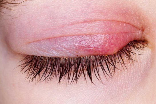
Corneal staining. There is no specific ocular surface finding that indicates asymptomatic dry eye — the only indication is that the patient does not have symptoms, yet the physician sees something on the ocular surface: an increase in tear evaluation, a decrease tear production, staining, and so on.
She also asks about environmental circumstances that could lead to rapid evaporation of tears, such as sleeping under a ceiling fan, directing air vents towards the face, or even using a C-Pap unit for sleep apnea.
When a terrible ocular surface contradicts the patient’s claim of “feeling fine,” Dr. Galor looks for diabetic neuropathy and corneal nerve status. She will ask about chronic eyedrop use, because the common preservative, benzalkonium chloride (BAK), contributes to nerve damage.2 She also performs clinical tests to grade corneal sensation, or any difference between the patient’s eyes or both. She will also conduct confocal microscopy to evaluate nerve parameters, including density and tortuosity.
The risks of missing DED
One diagnostic challenge is that hyperosmolar eyes have greater variability in keratometry readings and IOL power calculations when compared with normal osmolar patients, according to a study by Dr. Epitropoulos, Dr. Matossian and others published in the Journal of Cataract & Refractive Surgery.3
Dry eye has traditionally been viewed as ocular discomfort, but there’s much more to it, says Dr. Akpek. She contributed to research published in the British Journal of Ophthalmology that correlates DED with a decrease in reading speed that could be attributable to reduced vision quality. “Particularly reading or performing computer work for prolonged periods of times, those are difficult for dry eye patients,” she says.
So what is the trajectory for asymptomatic patients who continue to go undiagnosed? Eventually, as the ocular surface becomes further damaged, common lifestyle activities such as wearing contact lenses, driving, or using electronic gadgets can be curtailed.
Obstructed meibomian glands may dilate, truncate and dropout, says Dr. Pepose. “As a result, there is often a reduced tear break-up time and often the vision fluctuates and the patient is not satisfied with the quality of vision. Ongoing punctate epithelial erosions will cause corneal haze, which translates into vision decrease. Corneal ulcerations or even perforation can occur in the most severe cases.”
Moving forward
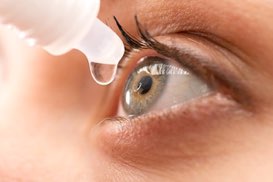
Once asymptomatic DED is identified, patients can start on a personalized treatment plan. Basic strategies to improve the ocular surface include a switch to preservative-free artificial tears, environmental modifications, and cutting back on anti-allergy medications, including topicals, that are drying to the ocular surface.
Better meibomian gland function is supported with oral omega-3 supplements, which provide the fatty acids, specifically eicosapentaenoic acid (EPA) and docosahexaenoic acid (DHA), naturally found in wild fatty fish.
The compliance conundrum
When patients feel no discomfort, how do you get them to comply with the doctor-prescribed therapy?
“I believe there will be adherence,” says Dr. Matossian, “if the prescribed treatment is explained clearly and if the patient is made to understand the ‘why’ behind the treatment.” The patient has to have a clear understanding that DED is progressive to ensure compliance with the prescribed regimen.
Many clinicians share this philosophy. Dr. Pepose will share osmolarity scores with patients, to demonstrate the effectiveness of treatment. Dr. Epitropoulos educates patients using tools such as meibography to demonstrate that MGD is a progressive disease and if not treated may lead to atrophy and irreversible damage to the glands. “DED can also delay healing and affect the outcomes of our surgical procedures, so this often motivates patients to be compliant with treatment.” OM
Esen K. Akpek, MD, has received institutional research grants from Allergan.
Alice Epitropoulos, MD, FACS, is a consultant to Allergan, AMO, BioTissue, TearScience, Bausch + Lomb, NovaBay, Omeros, Physician Recommended Nutriceuticals, RPS, Shire Pharmaceuticals and Tear Lab Corporation. She has performed contracted research for PRN, Ocular Therapeutix, Kala Pharmaceuticals, B + L and TearLab Corporation. Also, Dr. Epitropoulos holds property/patent rights for the EpiGlare Tester.
Anat Galor, MD, MSPH, reports no relevant financial disclosures.
Cynthia Matossian, MD, FACS, has financial relationships with Abbott Medical Optics, Alcon, Allergan, ALPHAEON, Bausch + Lomb, Bruder Checked-Up, Imprimis Pharmaceuticals, i-Optics, Lenstec, Marco, Ocular Therapeutix, Inc., OMEROS, Physician Recommended Nutriceuticals, Progressive Tech Training, RPS Diagnostics, Shire, Strathspey Crown (Shareholder), Sun Pharmaceuticals, TearLab and TearScience.
Jay S. Pepose, MD, PhD, FARVO, is a consultant for Allergan, Abbott Medical Optics, Kala Pharma, Mimetogen Pharmaceuticals, MG Therapeutics, Shire and TearLab.
REFERENCES
1. Galor A, Feuer W, Lee D, et al. Depression, post-traumatic stress disorder, and dry eye syndrome: a study utilizing the national United States Veterans Affairs administrative database. Am J Ophthalmol. 2012;154:340-346.
2. Baudouin C, Labbé A, Liang H, Pauly A, Brignole-Baudouin F. Preservatives in eye drops: the good, the bad and the ugly. Prog Retin Eye Res. 2010;29:312-334.
3. Epitropoulos AT, Matossian C, et al. Effect of tear osmolarity on repeatability of keratometry for cataract surgery planning. J Cataract Refract Surg. 2015;41:1672-1677.
Ophthalmology Management, Volume: 20, Issue: August 2016, page(s): 60, 63, 64